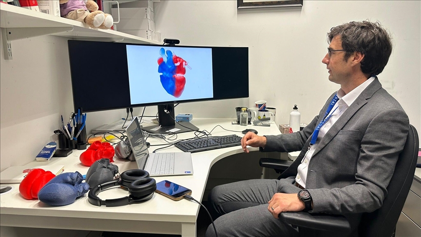
AI’s new beat: How digital heart ‘twins’ are redefining cardiac care
Researchers have created over 3,800 ‘digital twins’ of human hearts to study how cardiac disease varies across age groups, sexes, and lifestyles
 Imperial College’s Steven Niederer
- ‘We want to use these models to better understand how therapies might work in groups that were underrepresented in those trials, particularly women,’ Imperial College’s Steven Niederer tells Anadolu
LONDON
In a pioneering effort bridging the gap between engineering and medicine, researchers from King’s College London, Imperial College London, and The Alan Turing Institute have developed over 3,800 “digital twins” of human hearts, using artificial intelligence (AI) and real-world clinical data.
🫀 Researchers in the UK create over 3,800 digital models of human hearts, known as 'digital twins', to study cardiac diseases using artificial intelligence
💻 Using AI enables the researchers to create and analyse a vast number of patient-specific digital cardiac models on this… pic.twitter.com/j8GY5dlpQv
These computer-generated models, accurate down to the finest anatomical and physiological details, offer unprecedented insights into how heart disease varies across age groups, sexes, and lifestyles. The landmark research marks the first time such a vast number of patient-specific digital cardiac models have been created and analyzed at scale.
For the scientists behind the project, this achievement is more than just a technical milestone, signifying progress toward fairer, more personalized healthcare for millions.
“When we normally think about analyzing or interpreting medical images, we’re often doing that using rules built over many years,” Steven Niederer, chair in biomedical engineering at Imperial College and senior author of the study, told Anadolu.
“What we’re trying to do now is take ideas from how people design cars or airplanes – where you build a mathematical model and use it to predict performance – and apply that to the human heart.”
The groundbreaking research combines machine learning, clinical imaging, and advanced mathematical modeling techniques. An essential aspect involves training AI to automatically segment, or “label,” parts of medical scans to precisely identify heart structures.
“You might get an image which is just black and white, and we want to determine automatically which bits correspond to the heart,” explained Niederer.
Once labeled, the team constructs a mathematical mesh – a detailed 3D model simulating the appearance and function of each patient’s heart. These sophisticated models can run intricate simulations, illustrating how a patient’s heart responds to stress or particular treatments.
However, large-scale simulations are notoriously computationally intensive.
“We often have to send them away to big supercomputers, which might take hours to run a single simulation,” said Niederer.
“So, what we’re doing now is training AI models to approximate those simulations, cheaply and quickly, so we can personalize them to individuals much faster.”
Rethinking the rules of diagnosis
One of the most significant findings from this study challenges long-standing medical assumptions and highlights embedded biases, especially those relating to gender.
Traditionally, electrocardiograms (ECGs) have shown distinct differences between male and female patients. However, the digital twins revealed a surprising fact: these observed ECG differences might result more from structural variances than functional ones.
“We showed that ECG differences between men and women come down to size, not function,” said Niederer. “This challenges long-standing assumptions and may lead to more accurate diagnoses across sexes.”
Addressing such misconceptions is particularly vital in cardiology, where clinical trials have historically underrepresented women.
“We want to use these models to better understand how therapies might work in groups that were underrepresented in those trials, particularly women,” Niederer added. “Ultimately, it’s about making care more generalizable and more just.”
Digital twins in the real world
While much of this technology remains under development, the research team is already working to integrate it into real-world clinical settings.
Collaborating with hospitals in Nottingham and Sheffield and supported by The Alan Turing Institute, the scientists are building cloud-based software infrastructure that could one day plug directly into National Health Service (NHS) systems.
“We’re working on building software that allows you to take measurements from the hospital, send them into the cloud, automatically build your digital twin, and then send that back down to the clinicians,” Niederer explained.
“That’s how we make this technology real, by integrating it into actual clinical pathways.”
Currently, digital twins primarily assist in preparing for heart procedures. However, advances are being made to allow for daily, potentially continuous updates.
Researchers are exploring implantable sensors capable of providing real-time data – such as pressure and electrical activity – to continuously update digital twins, creating a dynamic model of a patient’s heart.
“This would allow for continuous monitoring and forecasting,” Niederer said. “It becomes a living model of your heart.”
Bridging science and humanity
Despite the sophisticated computational nature of the research, its greatest value lies in its direct connection to human health and quality of life.
“So, before being a treatment, before being a surgery, before trying to find out how to help a specific patient, the digital twins will be an extra tool to help these patients,” said Cristobal Rodero Gomez of the National Heart and Lung Institute at Imperial.
He emphasized the importance of anchoring technological developments in genuine human needs and realities.
“In this type of engineering project, it’s easy to get lost in the technical details,” he said.
“That’s why we speak with patients and the public, to keep asking: Why are we doing this? Who are we doing this for?”
This essential connection to patients’ experiences and realities is also embodied in one of the project’s most tangible outputs: 3D-printed hearts generated directly from digital twin models.
These physical representations serve multiple purposes, including education, patient communication, and even surgical planning.
“We find that this is a really great way to explain the work,” said Niederer.
“It makes what is otherwise a very computational representation of the heart something physical – something people can hold on to.”
The research team’s pioneering efforts have already extended beyond cardiology. Currently, the scientists are collaborating with Cancer Research UK to apply the digital twin approach to brain tumors, aiming to enhance treatment accuracy and efficacy.
The broader vision, according to the researchers, is to eventually create comprehensive, whole-body digital twins – responsive, predictive tools that can guide healthcare across multiple domains.
Anadolu Agency website contains only a portion of the news stories offered to subscribers in the AA News Broadcasting System (HAS), and in summarized form. Please contact us for subscription options.


